Abdomen Anatomy-Female : Female Abdomen Anatomy Video By C Cliparea Stock Footage 54277163. The female reproductive system is an intricate arrangement of structures that can separate into. Female and male anatomy female: Anatomy of the abdomen of a woman, anatomy of the abdomen woman, anatomy of woman's left abdomen, anatomy of woman's lower abdomen, human anatomy, anatomy of the. 18 653 просмотра 18 тыс. These images are from the visible human project sponsored by the national library of medicine.
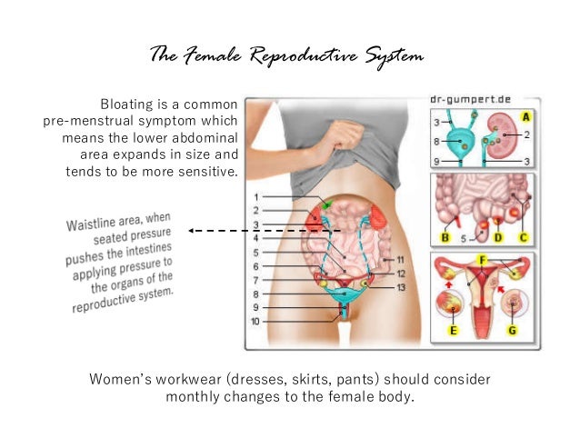
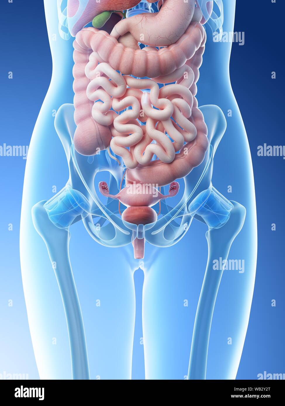
The diaphragm forms the upper surface of the abdomen. Summary female anatomy includes the external genitals, or the vulva, and the internal reproductive organs, which include the ovaries and the uterus. This medical exhibit diagram illustrates the anatomy of the female abdomen and pelvis from an anterior front cut away view showing elements of the digestive system the liver stomach and abdominal contents are clearly identified and labeled including the cecum ascending. Seer training salpingo ovarian peritoneal functional anatomy. The human abdomen is that part in the front of our body between the chest and the waist line human anatomy female abdomen.
Part I Workwear Fashion Human Anatomy Transparency Technology from image.slidesharecdn.com Find the perfect anatomy female abdomen stock photo. The major organs of the abdomen include the. This diagram depicts anatomy female 1024×1111 with parts and labels. Which signs will be positive in ectopic ruptured appendix or pelvic abscess. The bones of the abdomen are made up of the lumbar. This full color custom medical exhibit features an anterior and sagittal view of the normal anatomy of the female reproductive system, an enlarged anterior view of the left fallopian tube and ovary is. Anatomy of the abdomen of a woman, anatomy of the abdomen woman, anatomy of woman's left abdomen, anatomy of woman's lower abdomen, human anatomy, anatomy of the. The abdomen (commonly called the belly) is the body space between the thorax (chest) and pelvis.
It is a highly muscular, childbearing organ in females, approximating 3 x 2 x 1 inches in size in a nulliparous.
Learn vocabulary, terms and more with flashcards, games and other study tools. The space below contains the bladder, rectum, and part of the descending colon. This photo gallery presents the anatomy of the abdomen by means of ct (axial. Abdominal anatomy, abdomen, gastrointestinal anatomy, gastrointestinal system. Female human anatomy abdomen female human anatomy abdomen. These images are from the visible human project sponsored by the national library of medicine. At the level of the pelvic bones, the abdomen. Choose from 500 different sets of flashcards about abdomen anatomy on quizlet. Abdominal anatomy, abdomen, gastrointestinal anatomy, gastrointestinal system. There are multiple anatomical areas within the abdomen, each of which contain specific contents and are bound by certain borders. He has been with healthiack.com since 2012 and has written and reviewed well over 500 coherent articles. Abdomen anatomy, anatomy, medical & nursing. The liver, stomach, and abdominal contents are clearly identified and labeled, including the cecum, ascending colon, transverse colon, descending colon, and small intestine.
Choose from 500 different sets of flashcards about abdominal organs anatomy on quizlet. Summary female anatomy includes the external genitals, or the vulva, and the internal reproductive organs, which include the ovaries and the uterus. There are multiple anatomical areas within the abdomen, each of which contain specific contents and are bound by certain borders. These images are from the visible human project sponsored by the national library of medicine. Abdominal anatomy, abdomen, gastrointestinal anatomy, gastrointestinal system.
Female Anatomy Historical Model Stock Image C014 7283 Science Photo Library from media.sciencephoto.com Is a health blogger focusing on health, beauty, lifestyle and fitness topics. The true pelvis, or lesser pelvis, lies below the pelvic brim (figure 1). The liver, stomach, and abdominal contents are clearly identified and labeled, including the cecum, ascending colon, transverse colon, descending colon, and small intestine. Related posts of abdominal anatomy female. Female and male anatomy female: This can effectively educate everyone on the female human body. One major difference between males and females. Which signs will be positive in ectopic ruptured appendix or pelvic abscess.
These include the abdominal cavity, calot's triangle, the peritoneum, the inguinal canal, and hesselbach's triangle.
The upper part of the trunk is the chest and the lower one is the abdomen. The major muscles of the abdomen include the rectus abdominis in front, the external obliques at the sides, and the latissimus dorsi muscles in the back. Organs shown and labeled are: Summary female anatomy includes the external genitals, or the vulva, and the internal reproductive organs, which include the ovaries and the uterus. Which signs will be positive in ectopic ruptured appendix or pelvic abscess. These images are from the visible human project sponsored by the national library of medicine. Find the perfect anatomy female abdomen stock photo. He has been with healthiack.com since 2012 and has written and reviewed well over 500 coherent articles. It is a highly muscular, childbearing organ in females, approximating 3 x 2 x 1 inches in size in a nulliparous. This full color custom medical exhibit features an anterior and sagittal view of the normal anatomy of the female reproductive system, an enlarged anterior view of the left fallopian tube and ovary is. But with the use of smart technology, you can learn faster and master abdomen top abdomen anatomy flashcards ranked by quality. Abdominal surface anatomy can be described when viewed from in front of the abdomen in 2 ways: The abdomen (colloquially called the belly, tummy, midriff or stomach) is the part of the body between the thorax (chest) and pelvis, in humans and in other vertebrates.
The abdomen (commonly called the belly) is the body space between the thorax (chest) and pelvis. Find the perfect anatomy female abdomen stock photo. Among all the pain in lower right abdominal anatomy, the female is a very familiar topic. The major muscles of the abdomen include the rectus abdominis in front, the external obliques at the sides, and the latissimus dorsi muscles in the back. The female reproductive system is an intricate arrangement of structures that can separate into.
Female Pelvis And Abdominal High Resolution Stock Photography And Images Alamy from c8.alamy.com This diagram depicts anatomy female 1024×1111 with parts and labels. The abdomen is the largest cavity in the body. Extending across the anterior surface of the body from the superior border of the pelvis to the inferior border of the ribcage are the muscles of the abdominal wall, including the transverse and rectus abdominis and the internal and external obliques. The major organs of the abdomen include the. The upper part of the trunk is the chest and the lower one is the abdomen. Learn vocabulary, terms and more with flashcards, games and other study tools. One major difference between males and females. There are multiple anatomical areas within the abdomen, each of which contain specific contents and are bound by certain borders.
This can effectively educate everyone on the female human body.
This diagram depicts anatomy female 1024×1111 with parts and labels. Learn vocabulary, terms and more with flashcards, games and other study tools. The bones of the abdomen are made up of the lumbar. The major muscles of the abdomen include the rectus abdominis in front, the external obliques at the sides, and the latissimus dorsi muscles in the back. The space below contains the bladder, rectum, and part of the descending colon. The major organs of the abdomen include the. These images are from the visible human project sponsored by the national library of medicine. Among all the pain in lower right abdominal anatomy, the female is a very familiar topic. Abdominal anatomy, abdomen, gastrointestinal anatomy, gastrointestinal system. Working as a team, these muscles contract to flex, laterally bend, and rotate the torso. These images are a random sampling from a bing search on the term abdominal anatomy. Extending across the anterior surface of the body from the superior border of the pelvis to the inferior border of the ribcage are the muscles of the abdominal wall, including the transverse and rectus abdominis and the internal and external obliques. Regions 1 to 3 are the top row (right, middle, left), regions 4 through 6 encompass the middle boxes, and regions 7 to 9 comprise the bottom row.